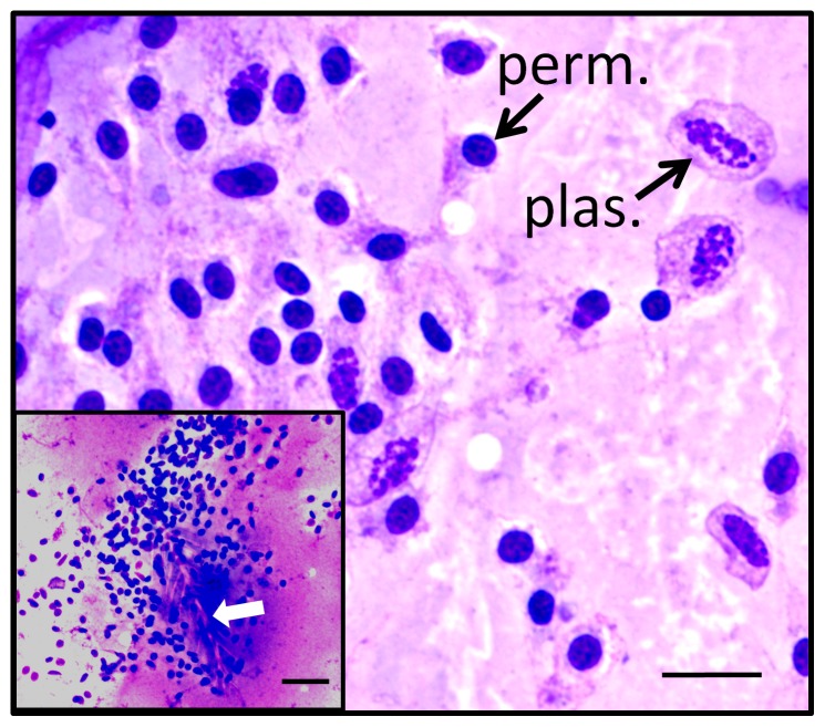
Figure 5. Aggregation of honey bee hemocytes involving permeabilized cells and plasmatocytes.
Hemocyte aggregates were occasionally found in Wright-stained smears of hemolymph samples inadvertently containing setae from the surface of the bee (white arrow, inset). Resulting aggregates included both plasmatocytes (plas.) and permeabilized cells (perm.). Plasma membranes were seen surrounding plasmatocytes. This observation contrasted with permeabilized cell nuclei that appeared to be associated with variable amounts of cytoplasm, with little evidence of plasma membranes. The total magnification for each image is 1000X, and the scale bar represents 10 µm for the primary image and 20 µm for the inset.

