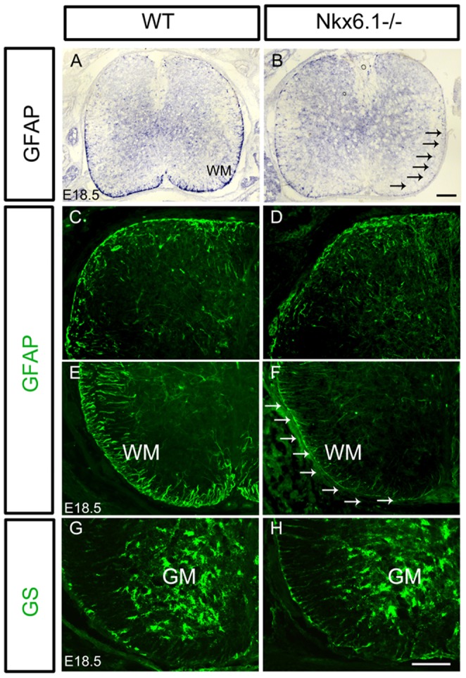
Figure 4. Defective differentiation of fibrous astrocytes in the ventral white matter of Nkx6.1 mutant spinal cord.

Spinal cord sections from E18.5 wild-type and Nkx6.1 mutants are subjected to ISH with GFAP riboprobes (A, B), or immunostaining with anti-GFAP (C-F) or anti-GS (G, H). In Nkx6.1 mutants, GFAP expression was reduced in the ventral, but not dorsal white matter. Strong GS signal was observed in the ventral gray matter of spinal cord, and no significant difference was detected between control and mutant tissues (G, H). Arrows indicate the reduced GFAP staining in the mutant tissues. Scale bars: 100 µm.
