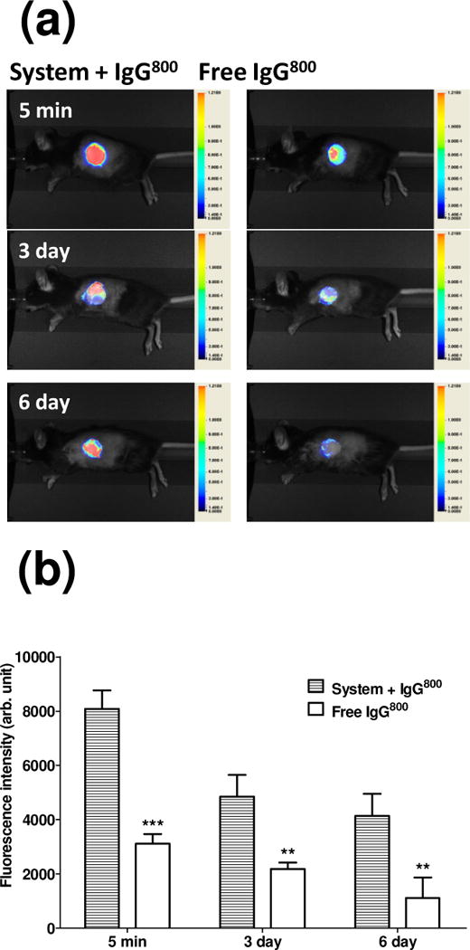
Figure 4.

Real-time monitoring of model antibody in graft bed in vivo. (a) Representative real time images of recipient C57BL/6 administrated with solution containing EAK16-II, EAKIIH6, αH6-IgG, pAG, and IgG800 (“System + IgG800”) or IgG800 alone in PBS (“Free IgG800”) at the graft bed prior to the skin graft. (b) Fluorescence at the graft site. Student’s t test was used for statistical analysis at each time point (n=3–6, ***: p < 0.001, **: p < 0.01).
