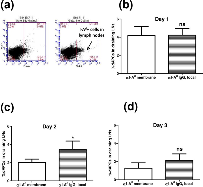
Figure 5.

Detection of I-Ad dAPCs in draining lymph nodes. αI-Ad was administrated at the graft bed with the self-assembling components (“αI-Ad membrane”) or PBS (“αI-Ad IgG, local”) prior to the skin graft. Single cell suspensions enriched for APCs were prepared from draining lymph nodes of recipient C57BL/6 mice. Cells were stained with Allophycocyanin conjugated anti-mouse MHC-II I-Ad antibody, and flow cytometry was used to probe the frequency of I-Ad dAPCs. (a) Representative dAPCs frequency on day 2. Student’s t test was used to confirm the statistical difference on day 1(b, n=4, ns: not significant), day 2 (c, n=3-, *: p < 0.05), and day 3 (d, n=3, ns: not significant).
