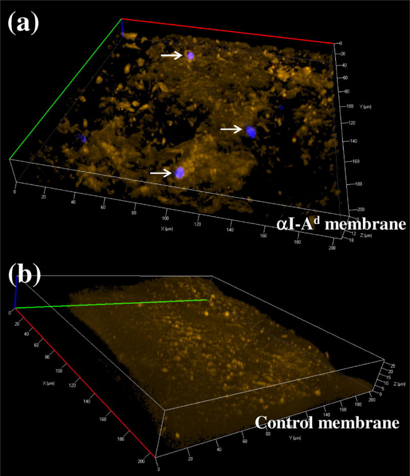
Figure 7.

Immobilization of dAPCs on membrane imaged using confocal microscopy (40×). dAPCs were induced to egress using CCL21. Cells adsorbed on membranes with αI-Ad (“αI-Ad membrane”) or without (“Control membrane”) were stained for DNA (visualized using a DAPI filter) and fixed. Cells ingested with nanoemulsions were illuminated with DiR (red). Membranes were imaged with a Cy3 filter set (orange).
