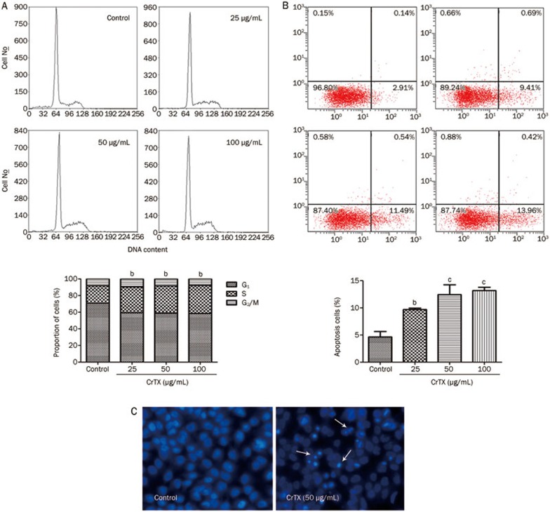
Figure 3.
Effects of CrTX on cell cycle and apoptosis of SK-MES-1 cells. (A) SK-MES-1 cells were seeded into 50-mL culture flasks and incubated with 0, 25, and 50 μg/mL, and 100 μg/mL CrTX for 48 h. At the end of the incubation period, cells were harvested and stained with PI. Cell cycle was analyzed by flow cytometry. (B) The percentage of apoptotic cells was measured by flow cytometry. (C) After treatment with CrTX for 48 h, cells were stained with Hoechst 33342 and morphology of apoptotic cells was observed under a fluorescence microscope (original magnification, ×200, Olympus, Tokyo, Japan). Condensed nuclei indicated cells underwent apoptosis.

