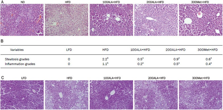
Figure 1.
Effect of ALA on histology analysis of liver tissue. (A) Representative hematoxylin and eosin (H&E) staining of a liver tissue sample (magnification 200×). (B) Liver sections of HFD-induced NAFLD were analyzed for steatosis and inflammation. bP<0.05 vs LFD. eP<0.05 vs HFD. (C) Representative periodic acid-Schiff (PAS) staining of liver tissue (magnification 200×).

