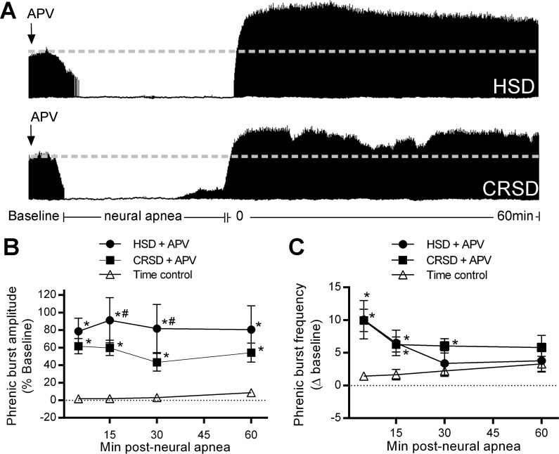
Fig. 2.
Spinal N-methyl-d-aspartate receptor (NMDAR) activation in CRSD rats, but not HSD rats constrains long-lasting iPMF. A: representative compressed phrenic neurograms depicting baseline, 30 min of neural apnea, and for 60 min after the resumption of phrenic neural activity in HSD and CRSD rats pretreated with intrathecal DL-2-amino-5-phosphonopentanoic acid (APV). Black arrows denote intrathecal delivery of APV. B: average phrenic burst amplitude (% baseline) for 60 min post neural apnea or an equivalent duration in time controls (triangles) in HSD (circles) and CRSD (squares) rats receiving APV prior to neural apnea. Both HSD and CRSD rats pretreated with APV and exposed to neural apnea expressed a significant increase compared with time controls at all time points, indicating long-lasting iPMF. This increase in CRSD rats was significantly lower than in HSD rats at 15 and 30 min post neural apnea. C: average phrenic burst frequency (Δ baseline) for 60 min post neural apnea or an equivalent duration in time controls (triangles) in HSD (circles) and CRSD (squares) rats pretreated with intrathecal APV prior to neural apnea. HSD rats receiving APV before neural apnea expressed a significant increase in phrenic burst frequency relative to time controls at 5 and 15 min. CRSD rats receiving intrathecal APV prior to neural apnea expressed a significant increase from time controls at 5, 15, and 30 min post neural apnea. Values are means ± SE. *Significantly different than time controls; #significantly different than HSD rats; P < 0.05.

