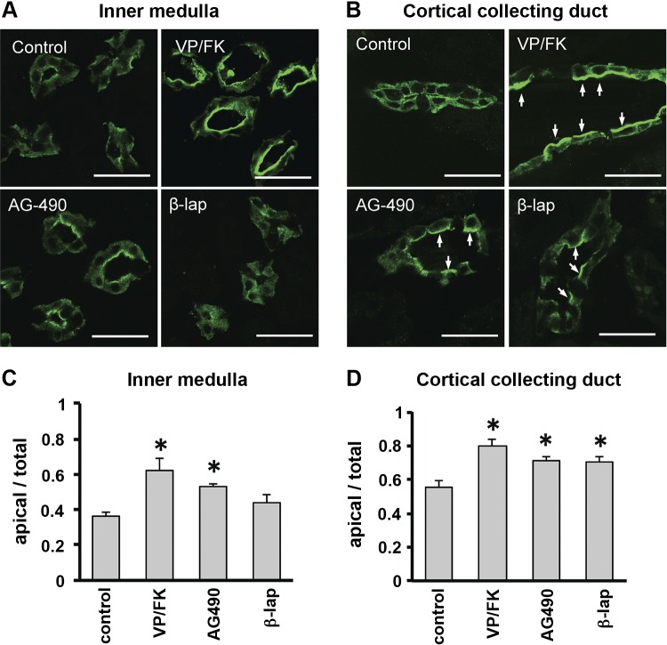
Fig. 4.
AQP2 accumulates on the apical plasma membrane in rat kidney slices after treatment with candidate compounds in vitro. Kidney slices from a Sprague-Dawley rat were incubated for 30 min with VP/FK (10 nM/10 μM), AG-490 (40 μM), or β-lapachone (50 μM). A: in inner medulla, AQP2 accumulated on apical membranes after VP/FK or AG-490 treatment. β-Lapachone had no detectable effect. B: in cortical collecting duct, AQP2 accumulated on apical membranes of principal cells (arrows) after VP/FK, AG-490, or β-lapachone treatment. Scale bars, 20 μm. C and D: quantitative analysis of AQP2 in inner medulla (C) and cortex (D). Fluorescence intensity of the apical area was divided by fluorescence of the whole cell area. Data are shown as means ± SE; n = 3. *P < 0.05 vs. control.

