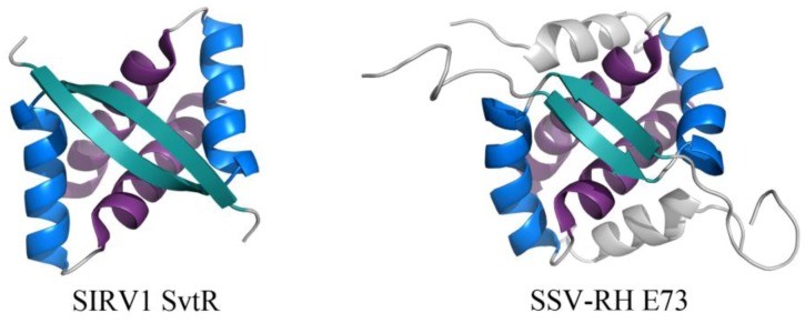
Figure 5.

RHH motifs in two unrelated proteins encoded by viruses from Fuselloviridae and Rudiviridae. The overall folds of SIRV1 SvtR (left, PDB ID 2KEL [33]) and SSV-RH E73 (right, PDB ID 4AAI [32]) are shown. The components of the RHH fold are highlighted for each dimer, including (in order of N- to C-terminal) β1 (turquoise), α1 (blue), and α2 (purple). SSV-RH E73 has an elaboration of the RHH fold, containing an additional α-helix (α3, colored gray) that may enhance the stability of its dimer and/or contribute to an additional ligand binding site.
