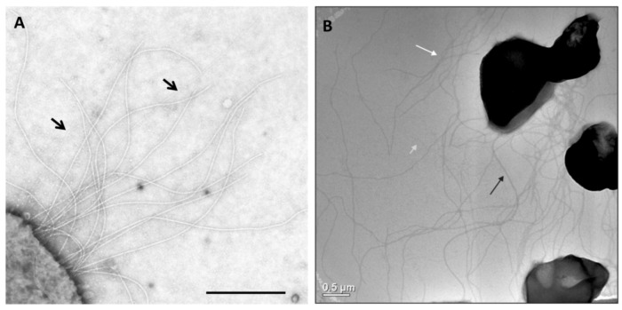
Figure 1.
Appendages on the well-studied Archaea M. maripaludis and S. acidocaldarius. (A) Electron micrograph of M. maripaludis showing thin pili (arrows) with thicker and more numerous archaella. Bar = 0.5 µm. Courtesy of S.I. Aizawa. Prefectural University of Hiroshima, Japan. (B) Electron micrograph of S. acidocaldarius showing the presence of three different appendages namely archaella (14nm diameter, black arrow), Aap pili (10–12 nm, white arrow) and threads (5 nm, grey arrow). Bar = 0.5 µm. Courtesy of A.-L. Henche and S.V. Albers, Max Planck Institute for Terrestrial Microbiology, Marburg Germany.

