
Figure 4.
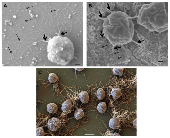
Role of archaella in attachment of Archaea to surfaces and other cells. Scanning electron micrograph of M. maripaludis attached to silicon wafer via thick cables of archaella (thick arrows) which can unwind to individual archaellar filaments (thin arrows). Bar = 100 nm (B). Connection of M. maripaludis cells to each other and underlying nickel EM grid via archaellar bundles. (A) and (B) reprinted from [31]. Bar = 100 nm (C). Scanning electron micrograph showing attachment of Mcc. villosus cells to a surface and to other cells via bundles of archaella. Bar = 1 µm. Courtesy of Gerhard Wanner, University of Munich, Germany.