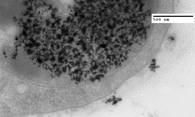
Figure 2.
TEM image of MTG-B tumor fixed in glutaraldehyde three hours post injection of 100 nm iron oxide nanoparticles into the tumor. Nanoparticles are seen both within and outside of the cell. Three hours appears approximately to be the time at which nanoparticles transition from being predominantly present outside of the cells to present within MTG-B cells.

