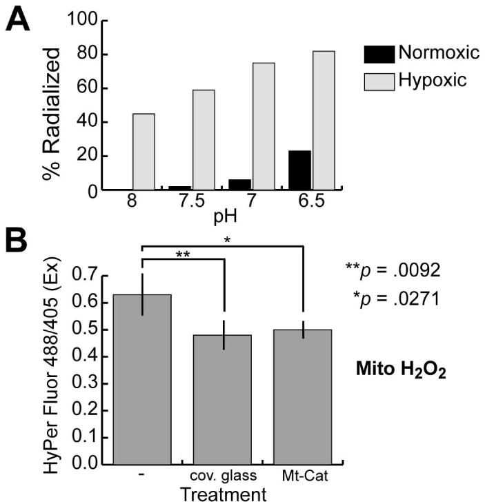
Figure 1.

Hypoxia induces radialized development and reduces mitochondrial H2O2 levels in sea urchin embryos. (A) Differential effects of controlled hypoxia and pH on development of oral-aboral polarity. Embryos were developed under the indicated conditions (see Supplemental Table 1 for details) until hatched blastula stage, then transferred to artificial seawater and developed to prism stage under normoxic conditions. At that point the embryos were scored for morphological radialization, defined as absence of ectodermal and endodermal asymmetry, and presence of multiple radially-arrayed spicules. The graph presents the combined results from the two experiments quantified in Supplemental Table 1. (B) Relative mitochondrial H2O2 levels in embryos subjected to the indicated treatments, measured using microinjected mRNA encoding HyPer-dMito (Evrogen), a mitochondrially-targeted yellow fluorescent protein derivative whose excitation maximum shifts from of 420 nm to 500 nm upon oxidation by H2O2. The indicated ratios were calculated from the average pixel intensities obtained from projected confocal fluorescence images of pre-hatching blastulae, obtained by exciting the embryos at 405 and 488 nm and collecting fluorescence emissions at 530 nm (three to six embryos imaged per treatment). The coverslip treatments were carried out as described in Coffman et al. (2004). Preparation and microinjection of mRNA and image analyses were performed as described in Coffman et al. (2009). Error bars depict the standard deviation; ANOVA followed by Dunnett's test was used to determine the indicated significance values (JMP version 8.0; Cary, NC). A second experiment using HyPer-cyto mRNA (Evrogen) gave similar results (Supplemental Fig. 3).
