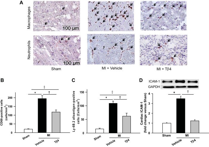
Fig. 2.
A: representative images of immunohistochemical staining for macrophages (top) and neutrophils (bottom) in the region of the infarct border at 7 days post-MI. B and C: quantitative analysis of CD68-positive cells (a marker for macrophage) and lymphocyte antigen (Ly) 6B.2-positive cells (a marker for neutrophils), respectively, showing that Tβ4 significantly reduced infiltrating macrophages and neutrophils. D: ICAM-1 (80 kDa) expression in the myocardium was significantly increased at 7 days post-MI, which was almost prevented by Tβ4. n = 5–7 animals/group. *P < 0.01; †P < 0.05; ‡P < 0.01.

