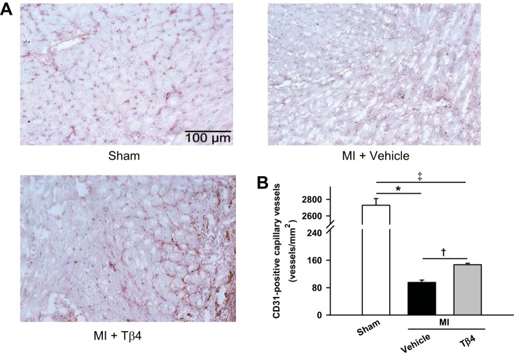
Fig. 4.
A: representative images of capillary vessels as indicated by positive staining for CD31 in the region of the infarct border. B: quantitative analysis of numbers of capillary vessels showing that capillary vessels were markedly reduced after MI but significantly increased with Tβ4 treatment. n = 5–6 animals/group. *P < 0.005; †P < 0.05; ‡P < 0.01.

