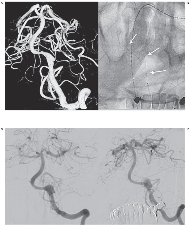
Figure 2.
A middle-aged man with SAH from a left vertebral dissection aneurysm (patient no.8). A) 3D angiogram shows a dissecting aneurysm in the V4 segment. Occlusion of this segment was not possible since the right vertebral artery had a PICA ending. B) Position of flow diverters. C,D) Follow-up angiograms after 2 (C) and 6 (D) months showing a slight enlargement of the dissecting aneurysm.

