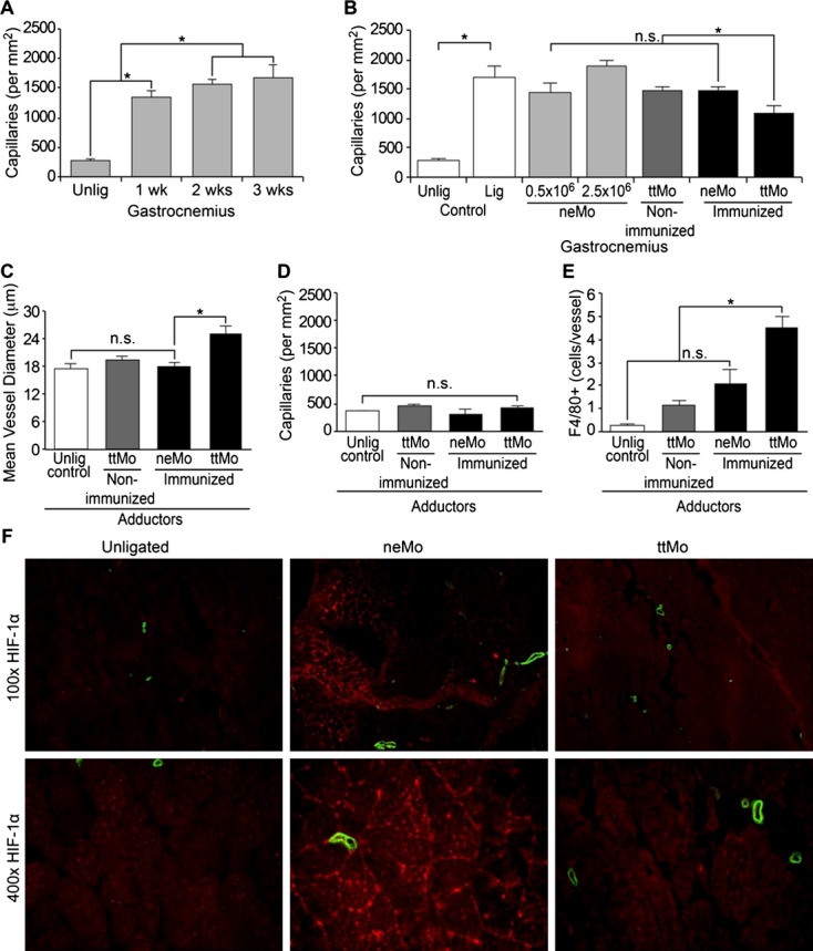
Figure 4.

A, Histologic workup of capillary density within the gastrocnemius muscle before and up to 3 weeks after ligation in untreated control animals with increasing capillary density after ligation (P=0.003, ANOVA); (B) Quantitative comparison of capillary density between the experimental groups 3 weeks after ligation; (C) Determination of mean vessel diameter within the adductor muscle, increasing vessel diameter in immunized mice after transplantation of ttMo (P=0.005, ANOVA; P<0.005, Tukey's post hoc test); (D) Quantitative analysis of capillary density in the adductor muscle (P=0.181, ANOVA); (E) Quantification of perivascular Mo infiltration after systemic cell transplantation (P=0.001, ANOVA); (F) Immunofluorescence staining of HIF‐1α (Cy3, red) and smooth muscle‐α‐actin (FITC, green) demonstrate low levels of HIF‐1α within the distal limbs of animals that had received ttMo, while normal saline controls and neMo transplantation exhibited pronounced HIF‐1α stabilization and high capillary density. ANOVA indicates analysis of variance; FITC, fluorescein isothiocyanate; HIF, hypoxia‐inducible factor; lig, ligated; neMo, non‐engineered monocytes; ttMo, tetanus toxoid monocytes; Unlig, unligated.
