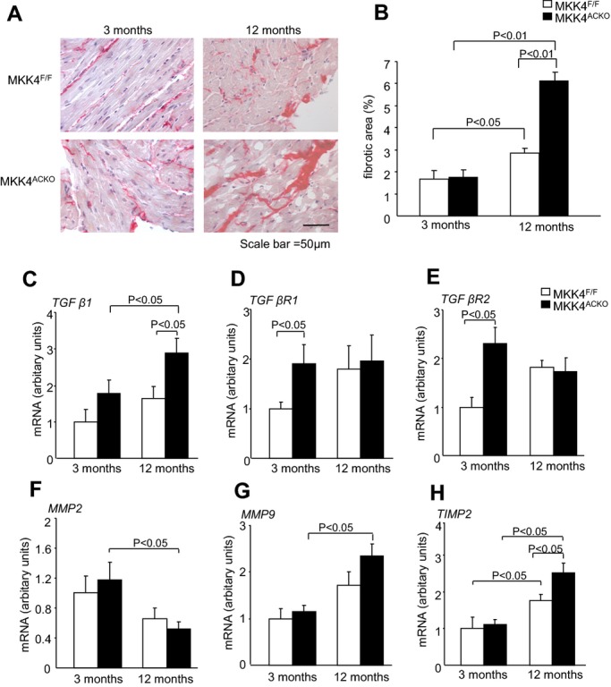
Figure 4.

Structural and molecular characterizations. A, Representative images of picrosirius red stained tissue, with fibrotic areas stained dark red, taken at 40× magnification in Mkk4F/F and Mkk4ACKO mice at ages of 3 (n=5) and 12 months (n=7). B, Quantification of the percentage area of fibrosis in young (left) and old (right), Mkk4F/F (white), and Mkk4ACKO (black) mice by picrosirius red staining. C through H, Real‐time PCR analysis of TGF‐β1 (C), TGF‐β receptor 1 (TGF‐β R1; D), TGF‐β receptor 2 (TGF‐β R2; E), MMP2 (F), MMP9 (G), and TIMP2 mRNA expression (H), from young and old Mkk4F/F and Mkk4ACKO left atria. The derived data are normalized to the housekeeping gene (HPRT) content, and the relative expression was calculated using the 2−ΔΔCT method. A Shapiro‐Wilk test was used to check whether data came from a normal distribution before data analysis using two‐way ANOVA followed by Bonferonni‐corrected post‐hoc t tests, with a value of P<0.05 considered to indicate statistical significance. Data are presented as means±SEMs (n=4 per group for both young and old Mkk4F/F and Mkk4ACKO). HPRT, hypoxanthine phosphoribosyltransferase; Mkk4ACKO indicates atrial cardiomyocyte specific mitogen‐activated protein kinase kinase 4 knockout mice; Mkk4F/F, Mkk4 flox/flox; MMP, matrix metalloproteinase; PCR, polymerase chain reaction; TGF‐β, transforming growth factor‐β; TIMP, tissue inhibitors metalloproteinase.
