
Figure 4.
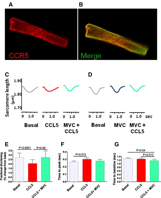
CCL5 decreases cardiomyocyte contractility via CCR5. Confocal microscopy showing expression of (A) CCR5 (red) on isolated adult rhesus macaque ventricular cardiomyocyte (RM VCM) merged with (B) sarcomeric actin (green). C, Representative twitch traces for VCM sequentially exposed to human recombinant CCL5 (100 nmol/L), then CCL5 (100 nmol/L) with maraviroc (MVC, 500 nmol/L). Compared to basal conditions (grey, left), CCL5 decreased contractility, shown by the reduced, flattened twitch amplitude (red, middle). The CCL5‐induced decline in contractility was reversed by subsequent addition of MVC with CCL5 (green, right). D, Representative twitch traces for reciprocal experiments showing VCM sequentially exposed to MVC (500 nmol/L) followed by a combination CCL5 (100 nmol/L) and MVC (500 nmol/L). Addition of MVC (blue, middle) did not alter contraction from basal (grey, left). Furthermore, addition of MVC prior to MVC+CCL5 (turquoise, left) prevented CCL5‐induced changes in contractility with twitch traces unchanged from basal. E, Summary data for RM VCM exposed to CCL5 (100 nmol/L) followed by MVC (500 nmol/L) with CCL5 (100 nmol/L). CCL5 significantly decreased fractional shortening compared to basal. Subsequent addition of MVC modulated the CCL5 effect towards basal shortening. F, CCL5 significantly increases the time from basal to 50% peak sarcomere length (t to pk) compared to basal conditions. Subsequent CCL5+MVC modulated t to pk shortening towards basal conditions. G, CCL5 did not significantly change the time from peak shortening to 50% baseline (t to bl) compared to basal conditions although t to bl in cells treated with CCL5+MVC was shorter than both basal and CCL5 treatment. Paired t‐tests, mean value indicated by the top of bar with bars representing standard error.