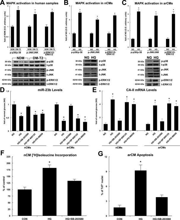
Figure 8.
MicroR‐23b (miR‐23b) downregulation is dependent on p38 MAPK pathway. A, p38 MAPK, ERK1/2, and JNK were significantly more phosphorylated in the myocardial samples from T2‐DM compared with NDM as shown by representative Western blots and O.D. semiquantitative analysis; *P<0.001 vs NDM, n=20 per group. B, High glucose (HG) significantly increased the phosphorylation levels of p38, ERK1/2, and JNK in nCMs in vitro. *P<0.001 vs normal glucose (NG), n=4 per group. C, HG significantly increased the phosphorylation levels of p38, ERK1/2, and JNK in arCMs in vitro. *P<0.0001 vs NG, n=4 per group. D, SB‐203580, PD‐98059, and SP‐600125 (p38, ERK1/2, and JNK specific inhibitors, respectively) effects on miR‐23b levels in high‐glucose (HG) treated nCMs or arCMs in vitro. *P<0.001 vs normal glucose (NG). E, SB‐203580, PD‐98059, and SP‐600125 effects on CA‐II mRNA levels in high‐glucose (HG)‐treated nCMs or arCMs in vitro. *P<0.03 vs normal glucose (NG), n=4 per group. F, SB‐203580 reduced HG‐induced nCM growth in vitro. *P<0.003 vs normal glucose (CON), n=4 per group. G, SB‐203580 reduced HG‐induced arCM apoptosis in vitro. *P<0.0008 vs normal glucose (CON), n=4 per group. Quantitative data are expressed as mean±SE. DM‐T2 indicates diabetes mellitus type 2; nCM, neonatal cardiomyocyte; arCM, adult ventricular cardiomyocyte; NDM, non–diabetes mellitus.

