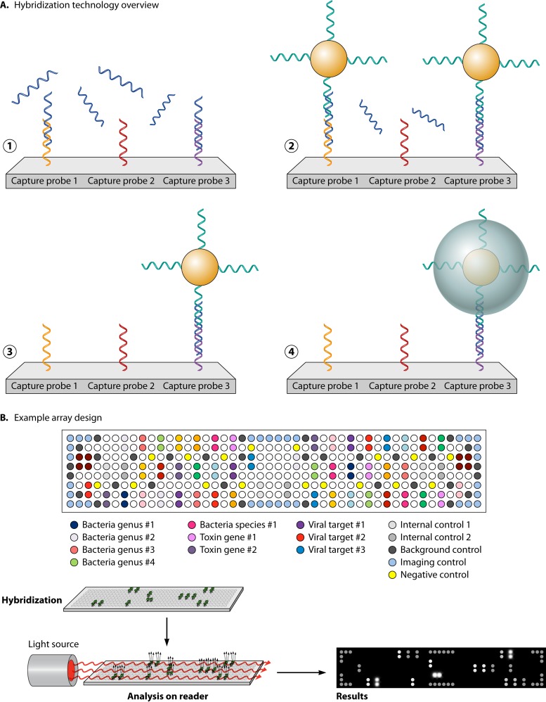
FIG 4.
Verigene solid-phase microarray. (A) Single-stranded, target-specific capture probes are arrayed spatially and immobilized onto the surface of a glass slide. The nucleic acid target (PCR amplicon or extracted nucleic acid) is denatured and applied to the glass slide. If present, the target nucleic acid will anneal to the complementary capture probe. Gold microspheres coated with single-stranded nucleic acid complementary to a different region of the target sequence are added and anneal to the capture probe-target sequence hybrid to form a “sandwich” nucleic acid structure. The array is washed to remove unbound nucleic acid and gold microparticles. Application of colloidal silver increases the size of the bound microspheres to increase the sensitivity of detection. (B) Target-specific capture probes, along with internal controls, are spotted in triplicate to different locations on the glass slide to ensure consistency of the annealing and hybridization steps and increase accuracy of results. Target detection is accomplished using a light source shown across the plane of the array. If present, bound silver microspheres diffract the light, which is then detected by an optical camera in the array reader.

