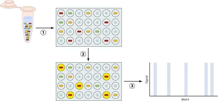
FIG 6.
Digital PCR. A nucleic acid template containing target sequences (colored boxes) in the original sample is diluted into individual microwells (plate PCR, pictured) or picoliter droplets (emulsion PCR) such that each well or droplet contains one or zero copies of the target sequence. Following partitioning of the specimen, endpoint PCR is carried out and amplicon is detected using fluorescent dyes or probes. Each well will be either positive or negative for fluorescent signal depending on the presence of the target sequence and resulting amplicon (yellow circles correspond to blue bars on the graph). The number of wells or droplets positive for fluorescent signal (yellow circles) directly corresponds to the number of specific target sequences (red boxes) present in the original sample.

