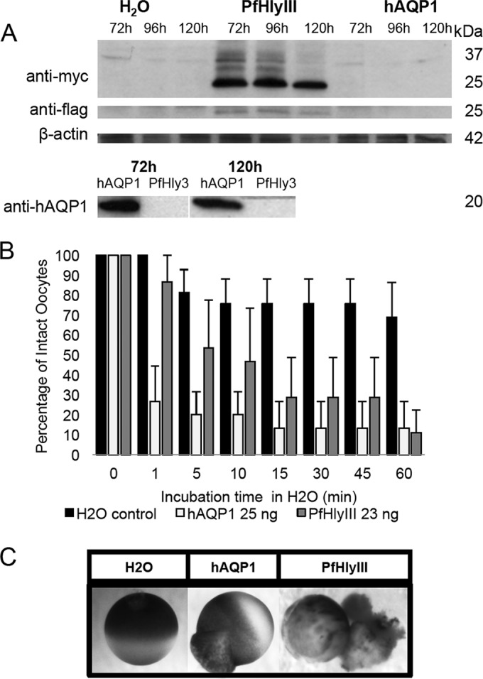
FIG 4.

Swelling and rupture of Xenopus oocytes expressing recPfHly III. (A) X. laevis oocytes injected with pXβG-ev1-myc-recPfHly III-flag cRNA express PfHly III (25 kDa), recognized by both anti-Myc and anti-Flag antibodies, whereas pXβG-ev1-hAQP1 cRNA-injected oocytes express hAQP1 (20 kDa). (B) hAQP1- and recPfHly III-expressing oocytes swell and rupture in a time-dependent manner in hypotonic buffer (water) at a higher rate than do the water-injected controls. Three independent experiments were conducted with three to six oocytes per group, and the mean percentage of intact oocytes was calculated and reported with the standard error of the mean. (C) Still photos of water control (45 min), hAQP1 (1 min), and PfHly III (5 min).
