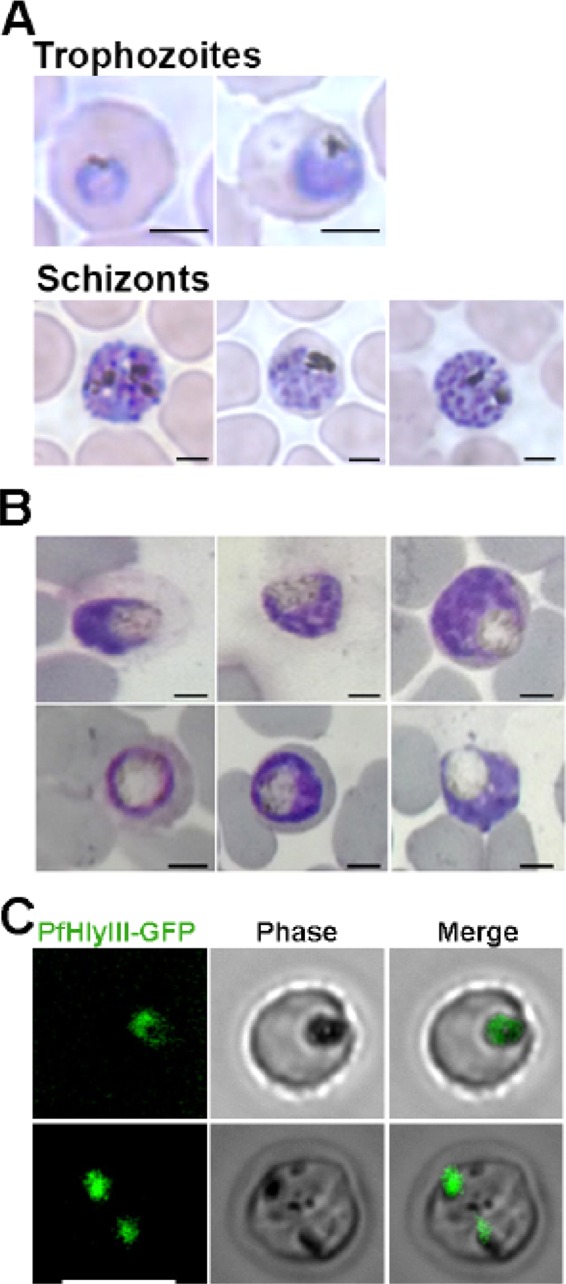
FIG 7.

PfHly III-GFP localizes to the digestive vacuole of P. falciparum. (A) Microscopy of Giemsa-stained nontransfected Dd2attB trophozoites and schizonts. (B) Giemsa-stained transfected P. falciparum with PfHly III-GFP fusion protein. Parasites demonstrate swollen digestive vacuoles. Bar, 2 μm. (C) Live fluorescence microscopy shows colocalization of PfHly III-GFP with hemozoin. Bar, 7 μm.
