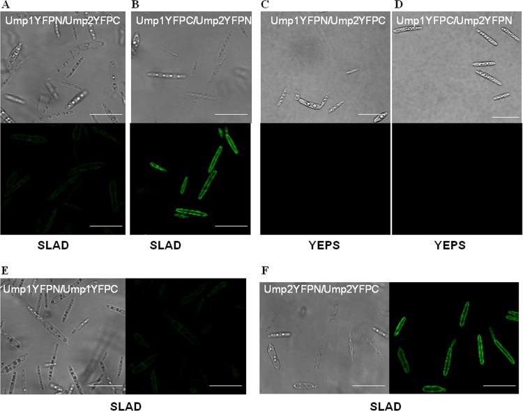
FIG 2.
Interaction between Ump1 and Ump2 under low-ammonium conditions. (A and B) Cells expressing Ump1 fused to the N terminus of YFP and Ump2 fused to the C terminus of YFP (A) or Ump1 fused to the C terminus of YFP and Ump2 fused to the N terminus of YFP (B) were grown for 24 h in liquid SLAD medium. (Top) Differential interference contrast images; (bottom) fluorescent images. (C and D) No fluorescence was detected for growth of the cells shown in panels A and B, respectively, on rich YEPS medium. (E and F) Differential interference contrast (left) and fluorescent (right) images of cells in which both the bait and the prey were either Ump1 (E) or Ump2 (F), grown on SLAD medium. Bars, 20 μm. For fluorescence results (and differential interference contrast images) for U. maydis strains in the ½ WT genetic background (40) expressing Ump1 or Ump2 fused to the N or C terminus of YFP, grown in SLAD medium, see Fig. S2 in the supplemental material (note the absence of fluorescence in each case).

