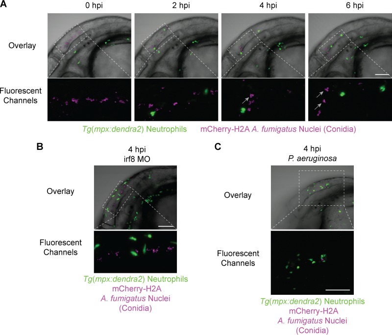
FIG 3.
Neutrophils do not phagocytose A. fumigatus conidia. Immunocompetent larvae with fluorescent neutrophils [Tg(mpx:dendra2)] were injected with A. fumigatus (gpdA::mCherry::his2A). (A) Time lapse imaging immediately following infection shows a lack of conidial phagocytosis by neutrophils (see also Movie S4 in the supplemental material). Gray arrows indicate conidial clumping resulting from macrophage phagocytosis. (B) Removal of macrophages with the irf8 morpholino (irf8 MO) leaves conidia in the extracellular space in the absence of macrophage phagocytosis (see gray arrows in panel A), and neutrophils remain nonphagocytic toward conidia (see also Movie S5 in the supplemental material). (C) Stimulated neutrophil recruitment with P. aeruginosa in a coinjection with A. fumigatus shows a continued lack of neutrophil phagocytosis of conidia. All images are representative of more than three experimental replicates. Scale bar, 100 μm.

