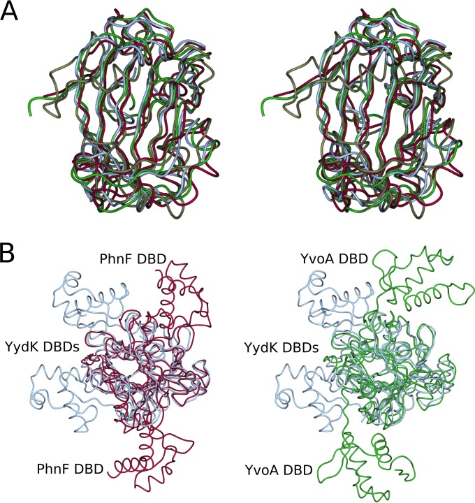
FIG 3.
Structural comparison of PhnF with related proteins. (A) Stereo image of the C-terminal domain of PhnF (purple) superposed with chorismate lyase (gray, PDB accession number 1G1B), B. subtilis YydK (blue, PDB accession number 3BWG), and B. subtilis YvoA (green, PDB accession number 2WVO). (B) Superpositions of YydK (blue, PDB accession number 3BWG) with YvoA (green, PDB accession number 2WVO) and PhnF (purple) looking down the β-sheet and showing the different positions of the N-terminal DNA binding domains (DBDs).

