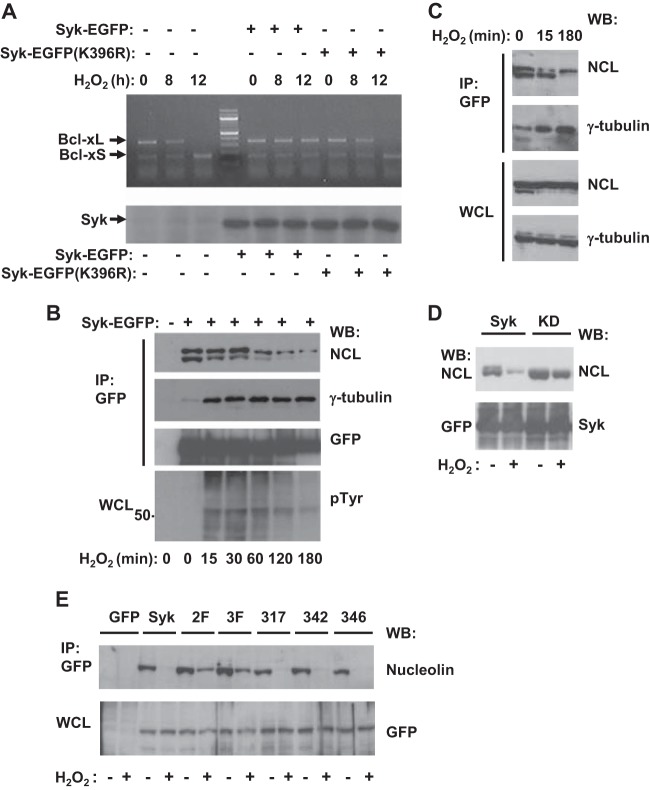
FIG 4.
Syk interacts with nucleolin. (A) MDA-MB-231 cells expressing rtTA but not Syk or MDA-MB-231 cells with Tet-regulated expression of Syk-EGFP or Syk-EGFP(K396R) (Lenti-X Tet-On) pretreated with doxycycline (+) were treated with 5 mM H2O2 for the indicated times. Cell lysates were analyzed by RT-PCR to measure the levels of Bcl-xL and Bcl-xS mRNA (top) or by Western blotting with anti-Syk antibodies to detect Syk-EGFP or Syk-EGFP(K396R) (bottom). (B) Tet-responsive MDA-MB-231 cells not induced (−) or induced with doxycycline to express Syk-EGFP (+) were treated with 5 mM H2O2 for the indicated times. Syk-EGFP was immunoprecipitated (IP) from cell lysates with GFP-nanotrap beads. Anti-GFP immune complexes were separated by SDS-PAGE and analyzed by Western blotting (WB) with antibodies against NCL, γ-tubulin, or GFP (to detect Syk-EGFP). Whole-cell lysates (WCL) were analyzed by Western blotting with antibodies against phosphotyrosine (pTyr) (bottom). The migration position of the 50-kDa molecular mass marker is indicated. (C) Syk-EGFP was immunoprecipitated with GFP-nanotrap beads from lysates of Tet-responsive MDA-MB-231 cells induced to express Syk-EGFP. Immune complexes and whole-cell lysates were separated by SDS-PAGE and analyzed by Western blotting with antibodies against NCL or γ-tubulin. (D) Proteins were immunoprecipitated with GFP-nanotrap beads from lysates of Tet-responsive MDA-MB-231 cells induced to express Syk-EGFP (Syk) or Syk-EGFP(K396R) (KD) and treated with (+) or without (−) 5 mM H2O2. Immune complexes and whole-cell lysates were separated by SDS-PAGE and analyzed by Western blotting with antibodies against NCL (top) and, to detect Syk-GFP, antibodies against GFP (bottom). (E) Proteins were immunoprecipitated with GFP-nanotrap beads from lysates of Tet-responsive MDA-MB-231 cells induced to express EGFP (lane GFP), Syk-EGFP (lane Syk), Syk-EGFP(Y342F/Y346F) (lane 2F), Syk-EGFP(Y317F/Y342F/Y346F) (lane 3F), Syk-EGFP(Y317F) (lane 317), Syk-EGFP(Y342F) (lane 342), or Syk-EGFP(Y346F) (lane 346) and treated with (+) or without (−) 5 mM H2O2. Immune complexes and whole-cell lysates were separated by SDS-PAGE and analyzed by Western blotting with antibodies against NCL (top) or GFP (bottom).

