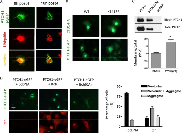
FIG 7.
Regulation of PTCH1 plasma membrane retention by K1413. (A) Colocalization of PTCH1-eGFP (green) and endogenous ubiquitin (red) in COS-1 cells at 6 h and 16 h posttransfection (post-t). (B) Subcellular localization of wild-type PTCH1-eGFP and HA-CTD1 and their K1413R mutants (green) shows increased plasma membrane levels of the K1413R mutants. (C) HEK293T cells expressing empty vector (pcDNA), PTCH1-HA, or the PTCH1 K1413R variant [PTCH1(KR)] were biotinylated as indicated in Materials and Methods. PTCH1-HA pulled down with avidin-labeled beads and total lysate PTCH1-HA were quantified by densitometry (right) after immunoblotting with an anti-HA antibody (left) (n = 3; *, P < 0.01). (D) (Left) Subcellular distribution of PTCH1-eGFP coexpressed in HEK293T cells with empty plasmid (pcDNA), myc-Itch, or catalytically inactive myc-Itch. (Right) Percentage of cells showing punctate staining (vesicular), perinuclear aggregates, or a mixed of the two. Values are means ± SEMs of 3 experiments.

