
FIG 4.
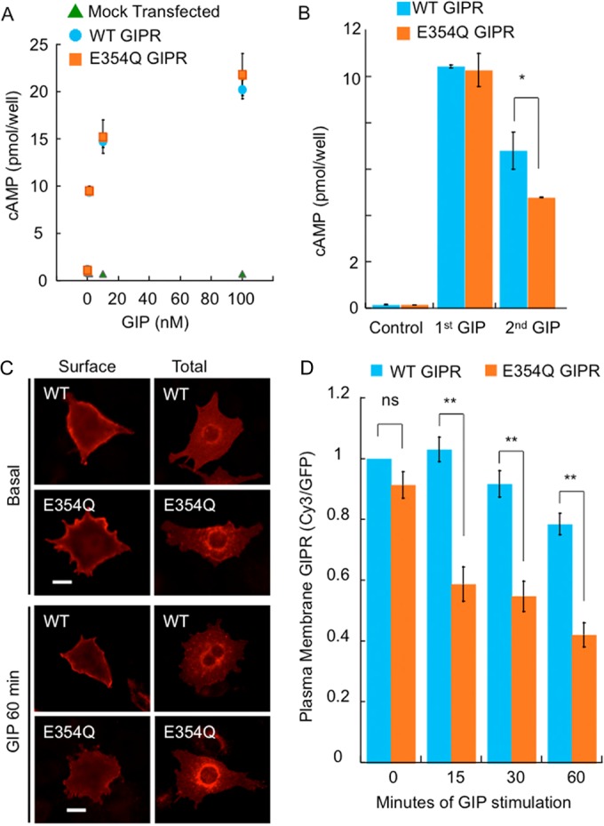
A natural variant of GIPR undergoes enhanced desensitization in response to GIP. (A) HEK293 cells transfected with either the empty vector, WT HA-GIPR-GFP, or E354Q HA-GIPR-GFP were stimulated with the indicated doses of GIP, and the amount of cAMP was measured. (B) HEK293 cells expressing either WT or E354Q HA-GIPR-GFP were incubated with or without 100 nM GIP for 1 h, followed by a second incubation with fresh GIP for 10 min. Cells were lysed, and the amount of cAMP was measured. (C) Adipocytes expressing WT or E354Q HA-GIPR-GFP were stimulated with 100 nM GIP for the indicated times, and anti-HA was revealed by indirect immunofluorescence of nonpermeabilized (Surface) and permeabilized (Total) cells. Representative cells are shown. Bar, 10 μm. (D) Quantification of cells treated as described above for panel C. The plasma membrane GIPR level was determined by indirect immunofluorescence of fixed, nonpermeabilized cells. The data from each experiment were normalized to the Cy3/GFP value for WT GIPR with no GIP treatment. Each bar is the average ± standard error of the mean from 3 to 5 independent experiments (**, P < 0.01; *, P < 0.05).