
FIG 9.
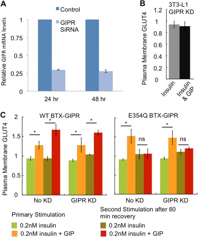
(A) Adipocytes were electroporated with GIPR siRNAs directed against the mouse GIPR sequence (CTAGGACAATCAACTGGAAGGC). After 24 and 48 h of electroporation, cells were harvested, and RNA was extracted. Quantitative PCR was performed as described in Materials and Methods. Each graph represents averages ± SD of data from 2 independent experiments. (B) The effects of 0.2 nM insulin and 0.2 nM insulin plus 100 nM GIP on the expression of HA-GLUT4-GFP in the plasma membrane were determined in 3T3-L1 adipocytes in which endogenous GIPR was transiently knocked down (KD). The knockdown of GIPR eliminated the effect of GIP on insulin-stimulated GLUT4 translocation. The data are averages ± SD of data from 2 experiments. (C) The amount of HA-GLUT4-GFP in the plasma membrane of adipocytes stably expressing WT BTX-GIPR or E354Q BTX-GIPR in which endogenous GIPR was transiently knocked down with an siRNA targeting mouse GIPR compared to cells in which endogenous GIPR was not knocked down was determined after a 30-min stimulation with 0.2 nM insulin or 0.2 nM insulin and 100 nM GIP (primary stimulation). In parallel, the amount of HA-GLUT4-GFP in the plasma membrane of adipocytes preincubated with 100 nM GIP for 60 min, washed free of GIP, and allowed to recover for 60 min before stimulation with either 0.2 nM insulin or 0.2 nM insulin and 100 nM GIP was determined (second stimulation). Each bar represents the average ± standard error of the mean of data from 3 independent experiments (*, P < 0.05; ns, not significant).