FIG 3.

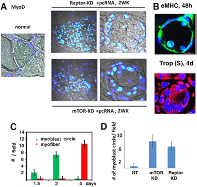
Myoblast rings in regenerating muscle. (A) DIC confocal images (gray) of sections of normal (left), Raptor-KD (upper right), or mTORC-KD (lower right) muscle after 2 weeks of pcRNA treatment, superimposed on merged DAPI (blue)- and anti-MyoD antibody (green)-stained nuclei. Left and right panels show lower and higher magnifications, respectively. (B) Ring structures in normal regenerating muscle after 2 days, stained for eMHC (green) or, after 4 days, stained for troponin (Trop) slow form (S) (red). Nuclei were DAPI stained. (C) Numbers of myoblast circles or myofibers per field (means ± SD) at various times in normal regenerating muscle. (D) Number of myoblast circles after 2 weeks in muscle expressing nontargeted (NT), mTORC-KD, or Raptor-KD muscle (means ± SD).
