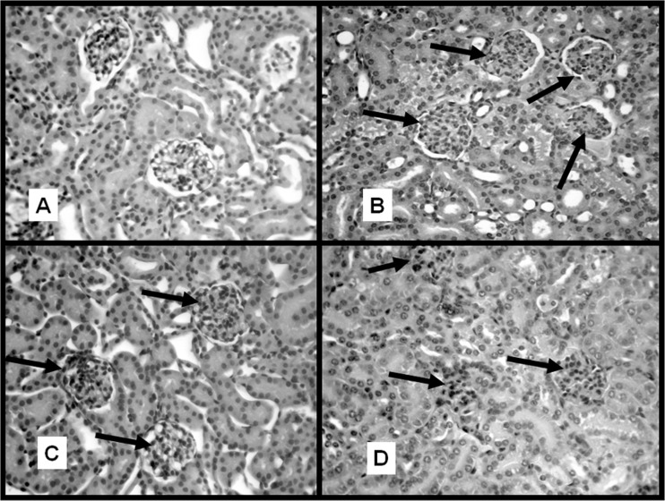
FIG 3.
Histological studies of kidney samples. Kidney samples from control and VA-D mice inoculated with PBS or EHEC were excised at 96 hpi, fixed, and stained with hematoxylin and eosin. Images were acquired with a Carl Zeiss III photomicroscope (Carl Zeiss AG, Oberkochen, Germany). Original magnification, ×250. (A) Kidney tissue of a noninfected control mouse with normal-aspect glomeruli and tubular epithelia preserved. (B) Kidney tissue of an EHEC-infected control mouse showing glomeruli (arrows) with hypercellularity, with some of the glomeruli retracted and the proximal and distal tubular epithelial cytoplasm showing a frosted-glass appearance and frayed luminal edges. (C) Kidney tissue of a noninfected VA-D mouse with normal-aspect glomeruli (arrows) and preserved tubular epithelia. (D) Kidney tissue of an EHEC-infected VA-D mouse showing glomeruli with marked shrinkage (arrow), mesangial hypercellularity, absence of Bowman's space (arrows), proximal and distal tubular epithelia with scant pale cytoplasm, and images of bare nuclei.

