FIG 4.
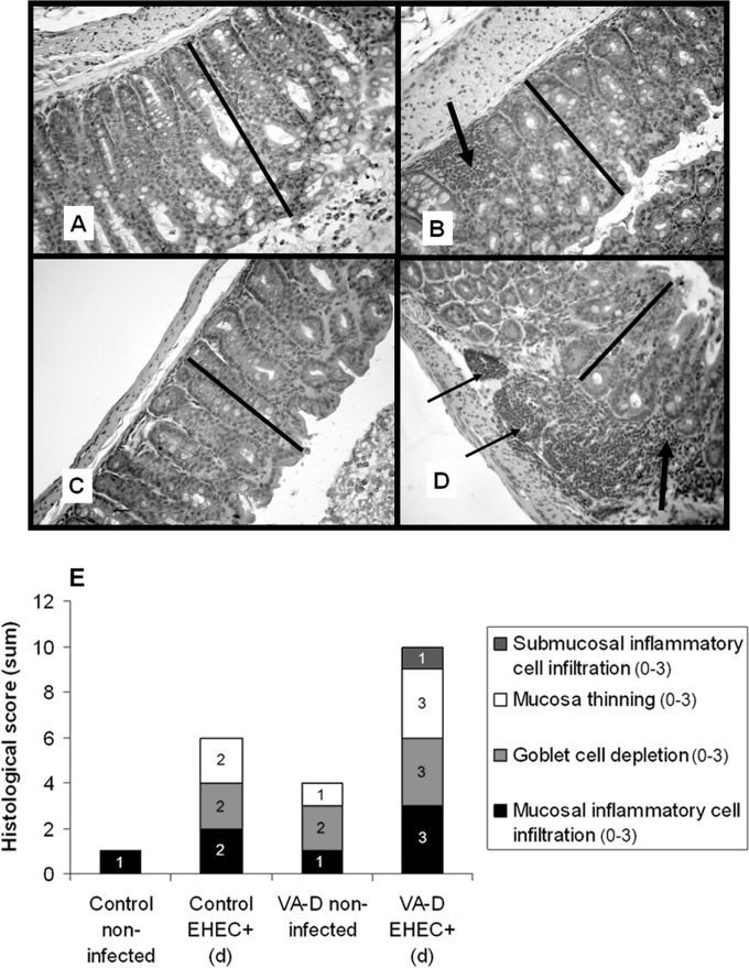
Histological studies of colon samples. Colon samples from control and VA-D mice inoculated with PBS or EHEC were excised at 96 hpi, fixed, and stained with hematoxylin and eosin. Images were acquired with a Carl Zeiss III photomicroscope (Carl Zeiss AG, Oberkochen, Germany). Original magnification, ×250. (A) Noninfected control mice show colonic mucosa with normal thickness (bar), preserved architecture, and abundant goblet cells. (B) EHEC-infected control mice show moderately thinned colonic mucosa (bar), a moderately decreased number of goblet cells, and lymphocyte inflammatory infiltrate in the lamina propria (arrow). (C) Noninfected VA-D mice show colonic mucosa with slightly reduced thickness (bar) and a decreased number of goblet cells with a mildly altered glandular architecture. (D) EHEC-infected VA-D mice show colonic mucosa with greatly reduced thickness (bar), architectural alterations, a sharp reduction in the number of goblet cells, and the presence of a chronic inflammatory infiltrate in the lamina propria (thick arrow) and submucosa (thin arrows). (E) Histological scoring of colon samples was performed in a blinded fashion by a pathologist. The y axis shows the sum of the histological scores for the following parameters: mucosal thinning (on a scale 0 to 3), goblet cell depletion (on a scale 0 to 3), mucosal inflammatory-cell infiltration (on a scale 0 to 3), and submucosal inflammatory-cell infiltration (on a scale 0 to 3). Three mice per experimental group were used.

