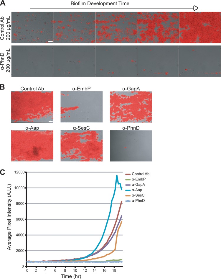
FIG 3.
Quantifying antibiofilm activity using a fluorescent reporter strain. (A) Longitudinal monitoring of S. epidermidis 1457-FL grown at 30°C in the presence of PhnD or control (rabbit anti-rat IgG) antibodies at 100 μg/ml. Images represent results of monitoring every 3 h at between 6 and 18 h of biofilm development. Scale bar, 20 μm. (B) Endpoint images at 20 h of 1457-FL biofilms grown at 30°C and treated with antibodies at 100 μg/ml. Scale bar, 30 μm. (C) Longitudinal quantification of the experiment described in the panel B legend using the average pixel intensity of images captured at 20-min intervals for 20 h. A.U., arbitrary units.

