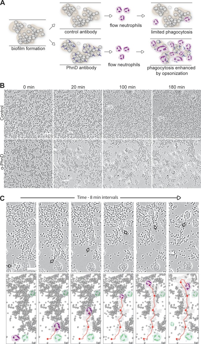
FIG 5.
PhnD antibodies enhance neutrophil phagocytosis of S. epidermidis biofilms. (A) Diagram of the opsonophagocytosis assay on preformed biofilms under conditions of flow. (B) Neutrophils exhibit increased binding, motility, and engulfment of biofilms in the presence of PhnD antibody versus the control (rabbit anti-rat IgG; both antibodies at 5 μg/ml). Images were taken at 0, 20, 100, and 180 min after introduction of neutrophils. Scale bar, 20 μm. (C) Tracking the antibiofilm activity of an individual neutrophil in the presence of PhnD antibodies. (Top) A single neutrophil (arrow) is capable of moving toward and engulfing large numbers of biofilm bacteria. (Bottom) Schematic representation of top panels to highlight the individual neutrophil (colored purple) and its path (red line) as it engulfs biofilm. Other neutrophils in the field of view are depicted in green. Scale bar, 10 μm.

