FIG 1.
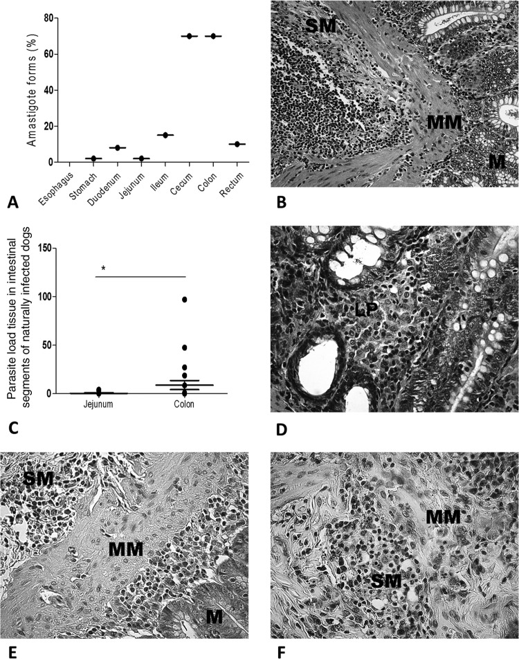
Parasite loads in dogs naturally infected with Leishmania infantum. (A) Frequencies of L. infantum amastigotes from the esophagus to the rectum. The cecum and colon harbored larger numbers of parasites than the other intestinal segments did. (B) Colonic tissue of an asymptomatic dog. The panoramic view shows the mucosa (M), muscularis mucosa (MM), and submucosa (SM). Note the intense and diffuse mononuclear cell infiltrate. Staining was done with HE. (C) Higher parasite loads in the colon than in the jejunum. (D) Higher-magnification view of panel B. Plasma cells predominate over lymphocytes and macrophages in the lamina propria (LP), without epithelial disruption. Staining was done with HE. (E) Immunolabeled amastigote forms of Leishmania throughout the mucosal (M), muscularis mucosal (MM), and submucosal (SM) layers (cecum-colon histological segment transition). (F) Higher-magnification view showing numerous immunolabeled parasites. Staining was done with streptavidin-peroxidase. *, P < 0.05.

