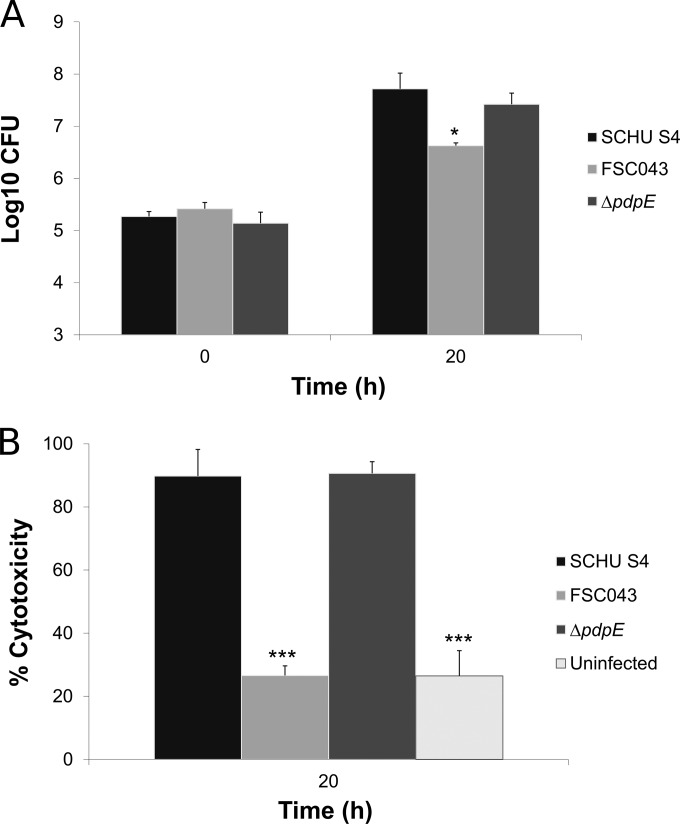
FIG 4.
Infection of J774 cells with the ΔpdpE mutant. (A) Cells were infected for 1 h with the indicated F. tularensis strains at an MOI of 30 and then incubated for 20 h. Bacterial replication was determined and expressed as mean log10 CFU of triplicate wells. Experiments were repeated twice with similar results. The asterisks indicate that the bacterial numbers were significantly different from the replication of the SCHU S4 strain at the 20-h time point. (*,P ≤ 0.05). (B) Culture supernatants of the infected J774 cells were assayed for LDH activity at the indicated time points, and the activity was expressed as a percentage of the level of noninfected lysed cells. Means and standard deviations of triplicate wells from one representative experiment of two are shown. The asterisks indicate that the cytotoxicity levels were significantly different from those of SCHU S4-infected cells for the 20-h time point, as determined by a t test (***, P ≤ 0.001).

