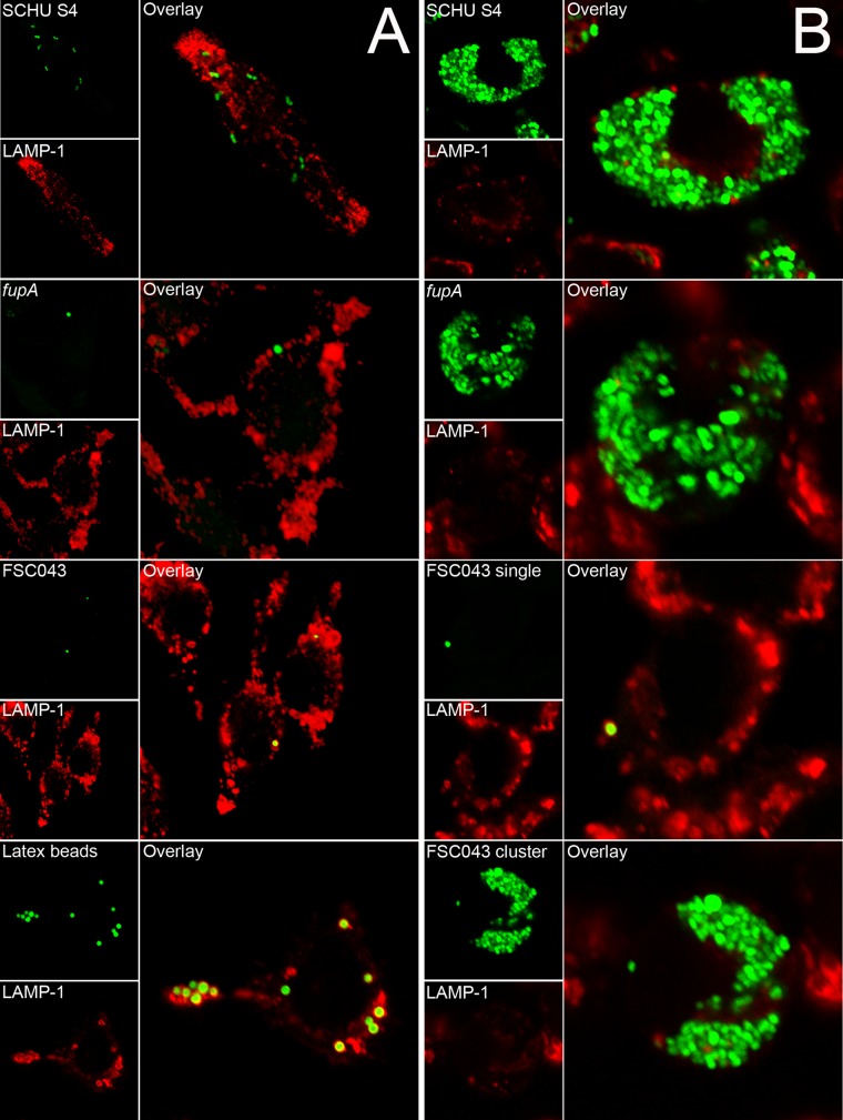
FIG 6.
Colocalization of GFP-expressing F. tularensis strains and the late endosomal marker LAMP-1. J774 cells were infected for 1 h with the indicated F. tularensis SCHU S4 strain at an MOI of 30 or latex beads at an MOI of 10 and further incubated for 3 h (A) or 18 h (B). In the representative confocal images, the green channel shows bacteria or latex particles and the red channel shows LAMP-1 staining for the indicated strain or latex beads. Confocal images were acquired with the Nikon C1 confocal microscope and assembled using Adobe Photoshop CS4 (Adobe Systems, San Jose, CA).

