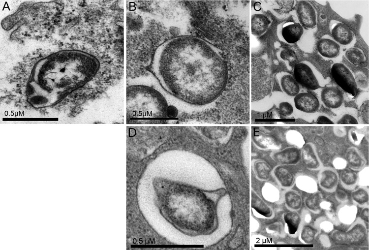
FIG 7.
Electron micrographs of J774 cells infected for 1 h with F. tularensis FSC043 or SCHU S4, the ΔiglC mutant, or the ΔiglC mutant and then further incubated for 18 h. (A) The ΔiglC strain. (B and C) A host cell containing an individual FSC043 bacterium enclosed by a phagosomal membrane (B) and a host cell containing a cluster of FSC043 bacteria without discernible phagosomal membranes (C). (D and E) A host cell containing an individual ΔpdpC mutant bacterium enclosed by a phagosomal membrane (D) and a host cell containing a cluster of ΔpdpC mutant bacteria without discernible phagosomal membranes (E). The electron micrographs were acquired with a JEOL 1200 EX-II electron microscope (JEOL Ltd., Tokyo, Japan) and assembled using Adobe Photoshop CS4.

