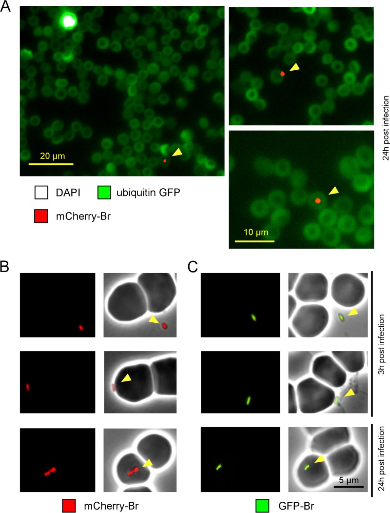
FIG 5.
Immunofluorescence microscopy of fixed or live bacteria. (A) Ubiquitin-GFP C57BL/6 mice were infected i.p. with 5 × 10⁷ CFU of mCherry-Br and bled at 3 h and 24 h p.i. The blood was fixed and then smeared on slides. The slides were dried overnight and then mounted in Fluoro-Gel medium containing DAPI nucleic acid stain (Electron Microscopy Sciences, Hatfield, PA) and examined under a fluorescence microscope. Brucella was found associated with erythrocytes mainly at 24 h, and thus we present only images starting at 24 h p.i. The figure shows examples of mCherry bacteria associated with DAPI- erythrocytes. The panels are color coded according to the antigen examined. Scale bar = 20 or 10 μm, as indicated. The data are representative of at least 3 independent experiments with a total of more 200 bacteria observed. (B and C) Wild-type C57BL/6 mice were infected with 5 × 10⁷ CFU of mCherry-Br (B) or GFP-Br (C). Blood samples were harvested at 3 h and 24 h p.i. and immediately diluted 10× in PBS. Freshly harvested blood samples were directly dropped on an agarose pad and sealed with VALAP before observation under a fluorescence microscope in a biosafety level III laboratory facility. The data are representative of at least 3 independent experiments, with a total of more than 30 bacteria observed under each condition.

