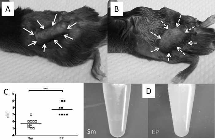
FIG 4.
(A and B) Appearance of S. mitis (A)- and EP (B)-infected chambers at 7 days after infection, indicated by arrows, showing swelling around EP-infected chambers. (C) Quantification of width of chamber tissue. (D) Appearance of fluid recovered from S. mitis-infected chambers (left) and EP-infected chambers (right). ***, P < 0.001 by t test.

