
FIG 5.
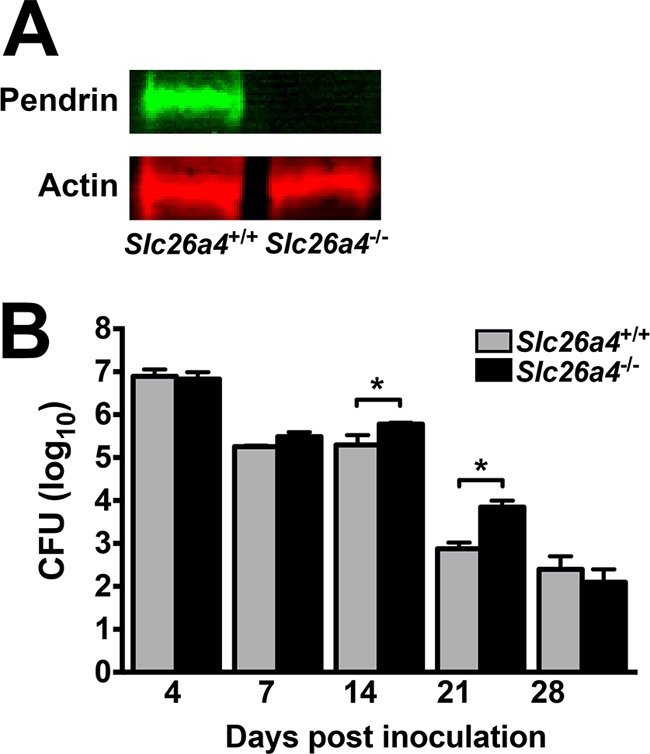
B. pertussis colonization is increased in Slc26a4−/− mice. (A) Lack of pendrin production (upper panel) in B. pertussis-infected Slc26a4−/− mice was confirmed by Western blotting, while actin levels (lower panel) remained normal. (B) Pendrin-expressing and pendrin KO littermate mice (n ≥ 5 per group) displayed a similar course of B. pertussis infection. However, at later time points, lung CFU were elevated in KO mice (black bars) compared with pendrin-expressing mice (gray bars). Bars represent the average per group ± SEM, *, P < 0.05.