FIG 3.
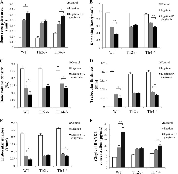
Bone resorption and gingival RANKL concentrations after ligature removal. Two weeks after treatment by ligation or ligation plus P. gingivalis infection, ligatures were removed from mice to allow wound healing for another 2 weeks. (A) The alveolar bone resorption around maxillary second molars (left and right) in WT, Tlr2−/−, and Tlr4−/− mice was measured and is presented as the area of bone loss (mm2). To further verify the observed difference in bone resorption between mice treated with ligation plus P. gingivalis and those treated with ligation alone, 3D bone assessment was performed using micro-CT to determine the remaining bone volume (mm3) (B), the bone volume density (% of bone surface/bone volume) (C), the trabecular thickness of remaining bone (mm) (D), and the trabecular number of remaining bone (1/mm) (E). (F) Total protein was isolated from gingival tissues, and RANKL production in gingival tissues was analyzed by ELISA using a specific antibody. Data are means and SE (n = 10). *, P < 0.05; **, P < 0.01.

