
FIG 5.
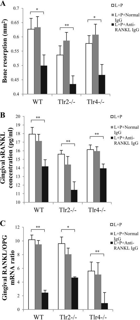
Bone resorption and gingival RANKL and OPG expression after RANKL blockade in WT, Tlr2−/−, and Tlr4−/− mice. Two weeks after treatment by ligation or ligation plus P. gingivalis infection, ligatures were removed from mice to allow wound healing for another 2 weeks. During this 2-week period, mice were injected with either rabbit anti-mouse RANKL IgG or normal rabbit IgG in the interdental papilla (1 μg/site) on both the buccal and lingual sides of the maxillary second molars (left and right), on days 15, 17, and 21. (A) At the termination of the experiment (day 28), the alveolar bone resorption around maxillary second molars (left and right) in WT, Tlr2−/−, and Tlr4−/− mice was measured and is presented as the area of bone loss (mm2). (B) Total protein was isolated from gingival tissues, and gingival RANKL concentrations were measured by ELISA. (C) Total RNA was isolated from gingival tissues, and RANKL mRNA expression and OPG mRNA expression in gingival tissues were detected by real-time PCR. The RANKL/OPG ratio was then calculated. Amplification of the GAPDH gene was used as a control. L, ligation; P, P. gingivalis infection. Data are means and SE (n = 10). *, P < 0.05; **, P < 0.01.