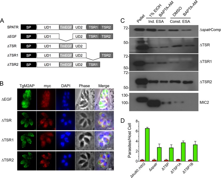
FIG 5.
TgSPATR domain deletion mutants are mislocalized. (A) Schematic illustrations of individual domain deletions of EGF, TSR, TSR1, and TSR2. (B) Indirect immunofluorescence localization of intracellular parasites using mouse anti-myc and rabbit anti-M2AP. (C) Induced (1% ethanol for 2 min) and constitutive (20 min) ESA fractions of all domain deletions. BAPTA-AM treatment for 10 min blocked the majority of secretion of a control MIC protein, TgMIC2, in both induced and constitutive secretion but not of the domain deletion mutants. Blots probed with mouse anti-myc or mouse anti-MIC2. (D) Red-green invasion assays of parasites after 20 min of incubation with HFF host cells. Parasites were stained as described in Materials and Methods. Data are means + SEM from two independent experiments, each with triplicate samples. The ΔEGF strain was not included in the secretion or invasion assays because it showed protein arrest in the ER.

