
FIG 4.
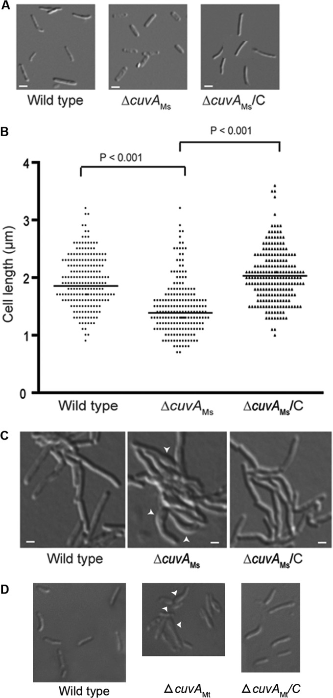
Morphology of M. smegmatis and M. tuberculosis cells. (A) M. smegmatis mc2155 (wild type), ΔcuvAMs, and complemented ΔcuvA (ΔcuvAMs/C) strains were grown in MM plus 0.01% cholesterol, harvested after 24 h, and examined by light microscopy. Bars = 1 μm. (B) Length distribution of M. smegmatis cells obtained after 24 h of growth in MM plus 0.01% cholesterol, determined by measuring over 200 cells of each strain and analyzed using ImageJ software. The horizontal lines indicate mean cell lengths. (C) Cells of each M. smegmatis strain were grown overnight on agarose pads made with MM plus 0.01% cholesterol and then photographed. (D) Wild-type M. tuberculosis H37Rv, ΔcuvAMt, and complemented ΔcuvA (ΔcuvAMt/C) strains were grown to late stationary phase in MM liquid medium plus 0.01% cholesterol and then examined by light microscopy using a 100× oil-immersion objective. Arrowheads in panels C and D indicate areas of abnormal cell morphology.