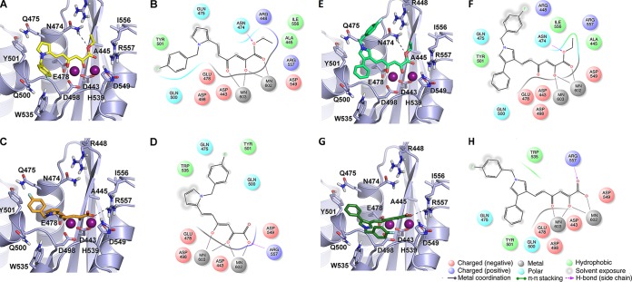
FIG 2.
Binding modes of DKA derivatives at the HIV-1 RNase H active site. (A, C, E, and G) Stick models of RDS1643 (A), RDS1644 (C), RDS1711 (E), and RDS1712 (G), represented as yellow, orange, light green, and dark green sticks, respectively. The receptor is shown as a gray cartoon. Amino acids involved in ligand binding are highlighted as sticks. The active site Mg2+ ions are represented as magenta spheres. (B, D, F, and H) Corresponding two-dimensional representations of DKA-RNase H interactions. Red and blue sticks represent oxygen and nitrogen atoms, respectively, and dashed lines represent hydrogen bonds.

