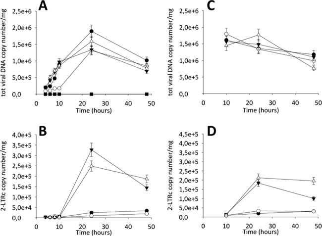
FIG 5.
qPCR kinetics of total and 2-LTRc DNA forms during a single round of HIV replication in the presence of inhibitors. MT4 cells were infected with HIV-1 in the absence (●) or in the presence of 10 μM RDS1759 (○), 10 μM RDS1760 (▼), 500 nM RAL (△), or 100 nM EFV (■), added at infection (A and B) or 10 h p.i. (C and D). Samples were analyzed for total viral DNA and 2-LTRc at different p.i. time points. The experiment was performed three times (error bars represent standard deviations). All samples were standardized and quantified relative to known standards, as described in Materials and Methods.

